Meniscus Transplant
The meniscus is used for transplant in the case of patients with knee joint pain and early joint degeneration, which appeared after the removal of the native damaged meniscus. This type of tissue, measured and processed, is engrafted into the patient via arthroscopic surgery and fixed inside the joint with stitches. In this way, it is possible to recreate the anatomy and biomechanics as close as possible to the original knee, with the final objective to prevent or at least slow down the arthrosis process.
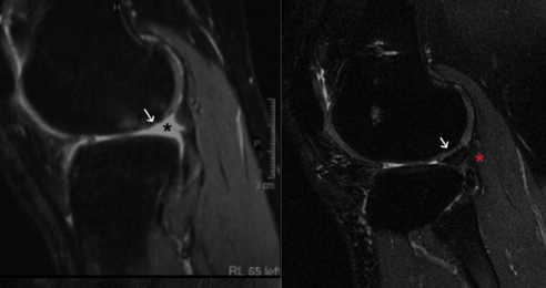
MRI of external meniscus transplant, on the left it is possible to notice the defect caused by a previous removal (black star). On the right, the defect is corrected by the transplanted meniscus (red star). Zaffagnini et al. Arthroscopy 2019
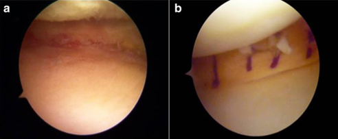
Arthroscopic Imaging of external meniscus transplant, on the left it is possible to notice the defect caused by a previous removal. On the right, the defect is corrected by the transplanted meniscus, interposed between the femur and tibia softening the load. Zaffagnini et al. JEO 2019
Ligament Reconstructions
Tendons from donors are a valid alternative to autologous tendons for knee joint ligament reconstructions. When a multi-ligamentous injury is present, or when a previous reconstruction was unsuccessful, often the patient’s tissues are not enough to guarantee an optimal reconstruction. In this case, the tendons from a donor, allow the reconstruction of the damaged ligaments without further damage to the anatomical structures of the patient (extensor apparatus, flexor muscles, contralateral intact knee). The good performance and safety of these tissues allow their application also in complex knee injuries such as chronic ruptures of the extensor apparatus, luxation of the kneecap, or tendons ingrained ruptures in patients with a knee replacement.
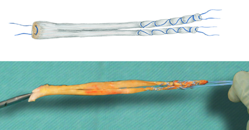
Achilles Tendon from donor used for knee joint ligament reconstructions. Zaffagnini et al. OTSR 2017
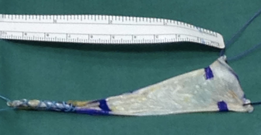
Fascia Lata from donor used for kneecap ligament reconstruction. Zaffagnini et al. KSSTA 2014
Tibial Osteotomy
The bone wedge from a donor is the most utilised tissue for filling the gap forming in the tibial metaphysis following an opening wedge osteotomy of the knee. Indeed, to correct alignment defects of the lower limbs that can also cause pain, it is necessary to cut the bone and position the two parts to create the correction required. Inserting a bone wedge between the gap favours the healing process of the cut and improves the postoperative recovery of the patient.
Fresh Joint Grafts
In patients with massive osteocartilaginous joint defects (knee, ankle), the use of massive joint transplants is a valid alternative in young patients with counterindication to standard prosthetics. Following proper procurement and conservation techniques, is possible to maintain the cell viability of the joint graft. The meticulous surgical technique can recreate the joint anatomy improving the quality of life in the patient and delaying the prosthetic replacement.