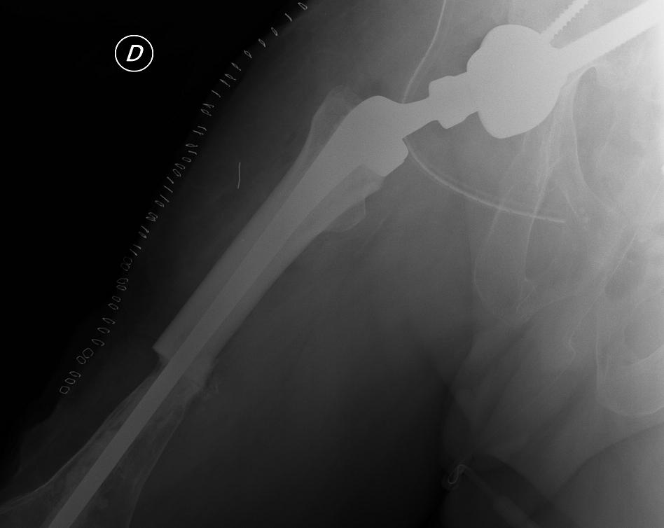
The use of musculoskeletal tissue is widely applied in prosthetic surgery, especially in prosthetic revision. A surgical revision of a prosthesis happens when the replacement presents several difficulties, mainly linked to the loss of bone tissue, a common consequence of repetitive surgical procedures. The lack of bone tissue is a fairly common consequence but also a very important problem, as they influence greatly the stability of the prosthesis in the bone, a very important parameter for successful replacement surgery.
When the prosthesis is not stable, the constant friction brings the wearing out of the bone around it (resorption, lysis). These losses of bone tissue require the application of specific grafts, generally of larger dimensions to increase the capacity of fixation and ultimately the stability of the replacement.
The homologous bone grafts most frequently used in replacement surgery are prepared with morselised bone and femoral heads that can be modified on the spot at the surgeon’s discretion. Ultimately, the application of bone shafts in periprosthetic fractures allows the reinforcement of the damaged bone and gives more strength to the fixation devices used to stabilise these kinds of fractures.
In the case of osteonecrosis and some cases of pseudoarthrosis, the following are used as filler grafts; lyophilised or demineralised bone chips or osteoinductive bone paste (Db-Graft, Db-Graft-T or I-Graft-C). Lyophilised bone must be rehydrated before use, also directly in the surgery room, adding autologous material such as bone marrow concentrate or platelet concentrate (PRP or PRF).
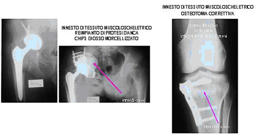
In the case of severe femoral bone defects that can occur both in revision and oncologic surgery, it is possible to use complex prosthetics. For example, in the case of a defect in the proximal femur, the surgeon proceeds to the full removal of the part and the defect is filled with a homologous massive graft in which the prosthetic stem is inserted (see image below). The advantage of such a technique is the customisation of the graft according to the bone defect and the maintenance of all the muscle insertion points on which the recipient’s muscles are sutured. In this way, the biomechanical properties of the joint and muscular strength are preserved.